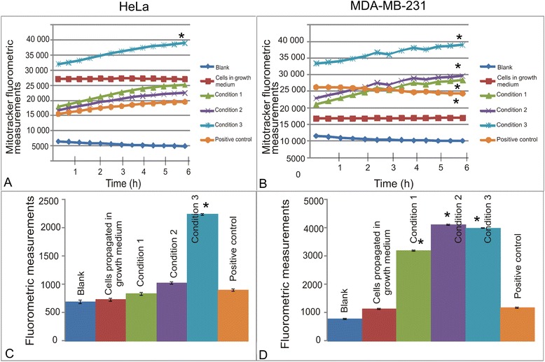
Fig. 4.

Mitochondrial membrane potential after partial- and complete glutamine- and glucose starvation for short term exposures and after 7 days. Mitochondrial membrane potential of HeLa cells (a) and MDA-MB-231 cells (b) exposed to medium consisting of varying metabolic conditions. Cells exposed to medium containing no glucose or l-glutamine were the only samples with reduced mitochondrial membrane potential indicating apoptosis induction. Fluorometrics and mitocapture demonstrated unsuccessful recovery and colony formation demonstrated that the HeLa (c) cell line was less prominently affected than the MDA-MB-231 (d) cell line. Little changes were observed in HeLa cells exposed to DMEM containing 3 mM-6 mM glucose and 0.5 mM-1 mM l-glutamine. However, cells exposed to medium containing 0 mM glucose and 0 mM L-glutamine demonstrated a statistically significant change in the mitochondrial membrane potential. With regard to the MDA-MB-231 cell lines, all three metabolic conditions affected the mitochondrial membrane potential cells exposed to DMEM containing 0 mM- 3 mM glucose and 0 mM-0.5 mM l-glutamine the most prominent. An asterisk (*) indicates P-value < 0.05
