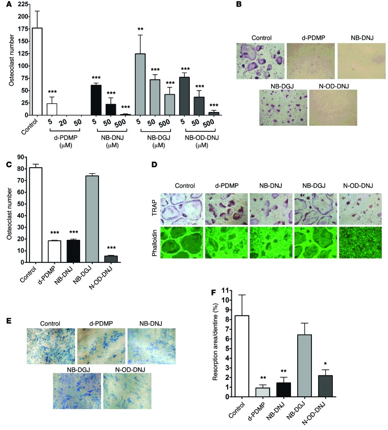
Figure 1. GSL inhibitors inhibit osteoclastogenesis and bone resorption in vitro.
(A) Mouse BM cells were cultured in the presence of 25 ng/ml M-CSF with 50 ng/ml RANKL (Control) and with or without d-PDMP (5, 20, or 50 µM), NB-DNJ, NB-DGJ, or N-OD-DNJ (5, 50, or 500 µM) in 48-well plates for 5 days, and TRAP-positive OCs were counted and shown as cumulative results from 3 different experiments. (B) Cultures with iminosugar inhibitors at 500 µM and d-PDMP at 20 µM were photographed (original magnification, ×40). (C and D) Mouse BM cells were cultured with 25 ng/ml M-CSF and 50 ng/ml RANKL in 48-well plates. On day 3 d-PDMP (20 µM) or NB-DNJ, NB-DGJ, or N-OD-DNJ (each at 500 µM) was added. Cells were cultured for another 48 hours before being fixed and stained for TRAP and counted (C) and phalloidin stained to identify mature OCs by the formation of actin rings (D) (original magnification, ×40). (E and F) Mouse BM cells cultured with M-CSF and RANKL on dentine slices with 5 µM d-PDMP or 50 µM iminosugars added on day 1 were evaluated on day 14 when resorption lacunae on dentine slices were visualized with methylene blue (original magnification, ×4). (F) Resorption area is expressed as a percentage of the dentine surface area. A representative experiment of three, each performed in triplicate assays, is shown for C and F. Data are mean ± SEM. Statistical analysis was performed using one-way ANOVA followed by Tukey’s multiple comparisons test (*P < 0.05, **P < 0.01, ***P < 0.001 versus control).

