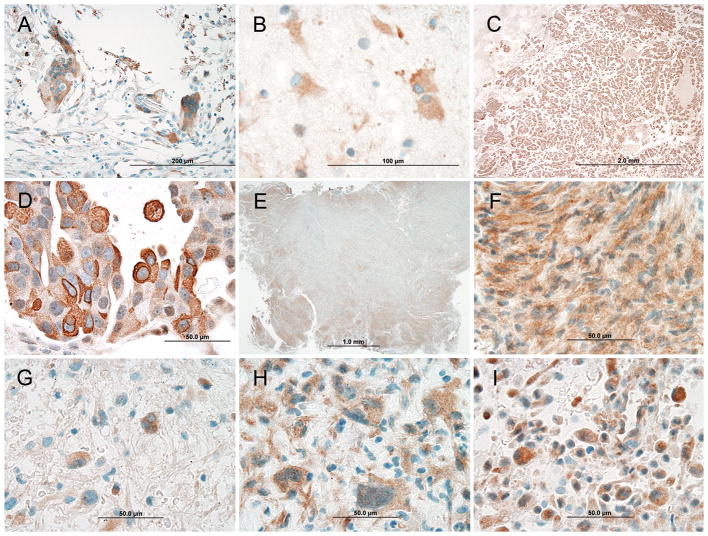
FIGURE 2.
Mammaglobin-A staining patterns of different central nervous system neoplasms. A and B, Weak (1+) to moderate (2+) intensity staining of stromal cells including multinucleated foreign body giant cells in a primary breast carcinoma and reactive astrocytes around metastatic carcinoma in the brain, respectively. C and D, Metastatic carcinoma showing diffuse architectural staining and moderate (2+) to strong (3+) cytoplasmic reactivity. E and F, Meningioma with patchy architectural staining and moderate (2+) cytoplasmic and nuclear staining. G–I, Three different glioblastomas showing moderate (2+) staining of the cytoplasm and nucleus.

