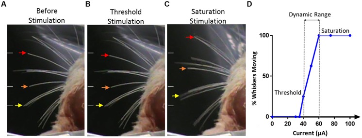
FIGURE 6.
Stimulation evoked whiskers movement. (A) The whiskers are in their initial positions before stimulation. Arrows indicate the movement of three whiskers of interest. White lines on the left of each panel indicate the initial position of the whiskers and are kept in the same position across all panels for reference. (B) Stimulation applied at threshold, only two whiskers, as identified by the orange and red arrows, were seen to move from their resting positions. (C) With saturated stimulation, many whiskers moved in either anterior or posterior direction from the resting points. Saturated stimulation also evoked whiskers movement on the ipsilateral side of stimulation which is not shown in this figure. (D) An example of the response from one electrode illustrating the percentage of whiskers moving versus current amplitude, threshold, saturation, and dynamic range are identified.

