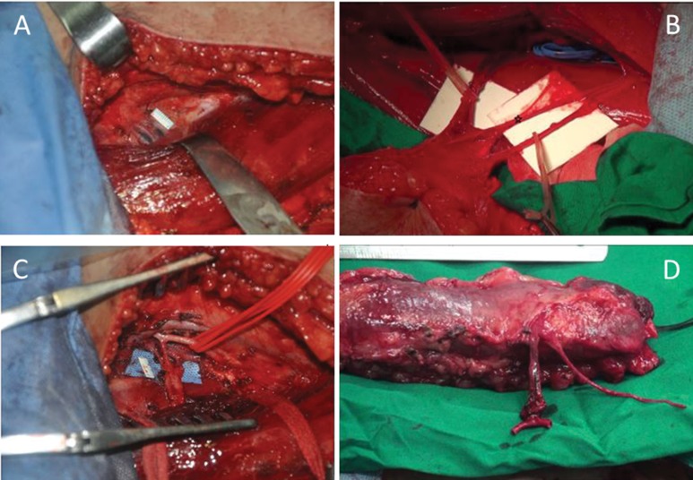
Figure 2.
Intraoperative images. A) Exposure of the dominant vascular pedicle. To avoid injury during the operation, the pedicle was not dissected at first. B) The neurovascular pedicle of the gracilis. Note that the sensory nerve branch (☆) must be resected to ensure enough motor nerve fiber regeneration (a, b). C) Exposure of the profunda femoris. A segment of the profunda femoris was prepared. It is unnecessary to perform a long dissection. D) The T-shaped arterial pedicle of the gracilis musculocutaneous flap (flap placed with the skin paddle downward).

