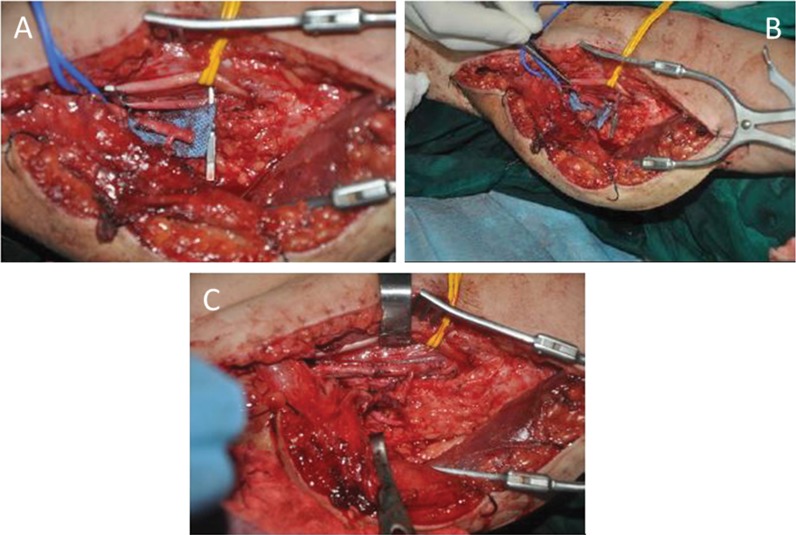
Figure 3.
Flow-through anastomosis of the T-shaped pedicle. A) The diameter of the profunda femoris segment is obviously larger than that of the nutrient artery of the gracilis. B) The brachial artery was resected, and the diameters of the segment profunda femoris and brachial artery were well matched. C) Interposed anastomosis to bridge the brachial artery. Two veins were anastomosed in direct end-to-end fashion.

