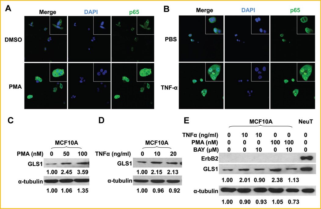
Fig. 5.
Activation of p65 is sufficient to upregulate GLS1 in MCF10A cells. A: MCF10A cells were treated with 100 nM of PMA in DMSO or DMSO alone for 6 h, followed by immunofluorescent staining for p65. B: MCF10A cells were treated with 20 ng/ml of TNF-α in PBS or PBS alone for 6 h, followed by immunofluorescent staining for p65. C: MCF10A cells were treated with 0, 50, or 100nM of PMA in DMSO for 6 h. GLS1 expression was determined by Western blots. D: MCF10A cells were treated with 0, 10, and 20 ng/ml of TNF-α for 6 h, GLS1 expression was determined by Western blots. E: MCF10A cells were treated with 10mMof BAY 11-7082 for 18 h, followed by treatment with 10 ng/ml of TNF-α or 100 nM of PMA for another 6 h. The whole cell lysates were collected for Western blotting analysis. The numbers under the blots are quantification of each detected bands; GLS1 or α-tubulin of control groups were set at 1.00, and treated groups were compared to the control group.

