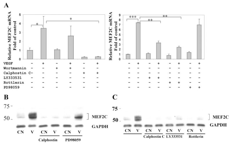
Figure 3.
Induction of MEF2C expression by VEGF is dependent on protein kinase C. (A) HRECs were pretreated with 100 nM wortmannin, 1 μM calphostin C, 2 μM LY333531, 5 μM rottlerin, and 50 μM PD98059 for 30 minutes, followed by treatment with VEGF (25 ng/mL) for 4 hours. Total RNA was isolated, and quantitative real-time PCR was used to measure the expression of MEF2C mRNA. Values were normalized to GAPDH gene expression. Results are representative of at least three independent experiments, each performed with duplicate samples. (B) HRECs were pretreated with calphostin C (1 μM) and PD98059 (50 μM) for 30 minutes, followed by treatment with VEGF (25 ng/mL) for 8 hours. Cells were harvested and probed with antibody to MEF2C. The same membrane was stripped and blotted with antibody to GAPDH for normalization. Results shown are representative of three independent experiments. (C) HRECs were pretreated with calphostin C, LY333531 (2 μM), and rottlerin (5 μM) for 30 minutes, then treated with VEGF (25 ng/mL) for 8 hours. Cells were then harvested for Western blot, as described. *P < 0.05, **P < 0.01, and ***P < 0.001 versus control.

