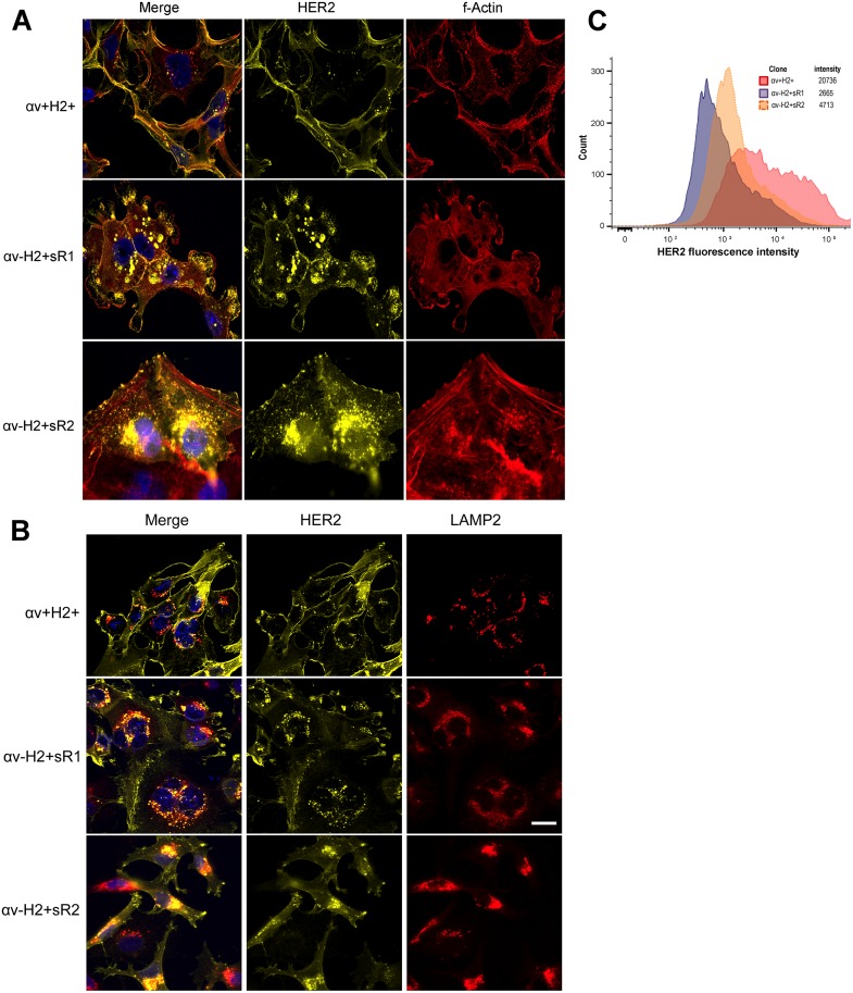
Fig 4. localization of HER2 in αv-integrin knockdown breast cancer cells.
MM2BH cell clones grown on glass coverslips were fixed, permeabilized and immunostained. (A) Deficiency of αv-integrin results in increased cytoplasmic localization of HER2 receptors. Yellow: HER2, using an antibody specific for the HER2 intracellular domain, red: f-actin, blue: nucleus. (B) HER2 co-localizes with lysosomes in αv-integrin knockdown cells. Yellow: HER2, red: lysosome-associated membrane protein2 (LAMP2), blue: nucleus. The images are representative of three independent experiments. Scale bar = 20μm. (C) Flow cytometry analysis of surface HER2 protein expression in live MM2BH clones.

