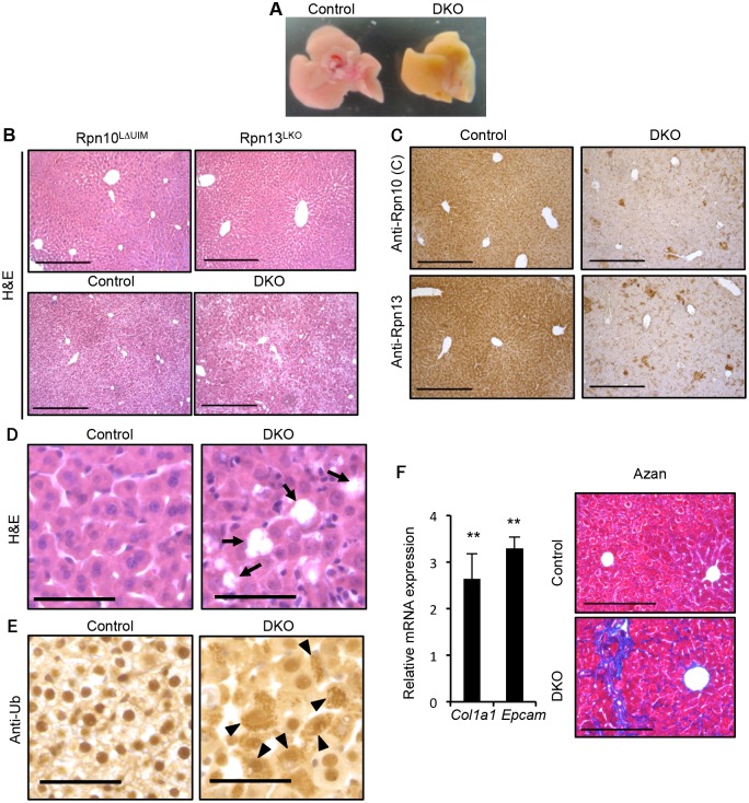
Fig 3. Simultaneous deletion of Rpn10-UIM and Rpn13 causes severe liver injury.
(A) Representative macroscopic images of 2-week-old control and DKO livers. (B) H&E staining of 4-week-old Rpn10LΔUIM, Rpn13LKO and 2-week-old control and DKO livers. All scale bars (black lines), 300 μm. (C) Immunohistochemical analysis on representative liver paraffin sections from 2-week-old mice by using Rpn10 (C) and Rpn13 antibodies. Scale bars, 300 μm. (D and E) Representative H&E staining (D) and immunohistochemical analysis of ubiquitin (E) on liver sections from 2-week-old mice. Arrows in (D) indicate regions of sloughing hepatocytes. Arrowheads in (E) indicate hepatocytes with high accumulation of ubiquitin in cytosol. All scale bars (black lines), 50 μm. (F) Azan staining of liver sections from 5-week-old control and DKO mice (right panels). All scale bars (black lines), 200 μm. Real-time RT-PCR was used to measure the expression of transcripts encoding fibrosis markers (Col1a1 and Epcam) in the livers of 3–6-week-old control and DKO mice (left panels). Data represent levels of transcripts in each genotype liver relative to those in control liver and are expressed as means; error bars denote SEM. **p < 0.01 (n = 4 each genotype).

