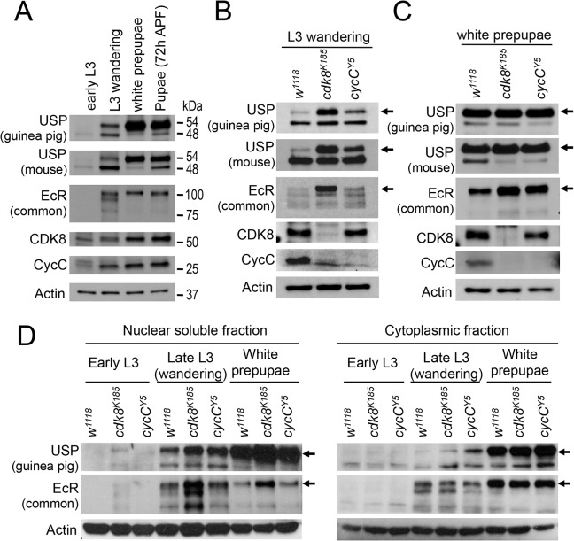
Fig 4. The levels and subcellular distribution of EcR and USP in cdk8 and cycC mutants in the third instar larvae and pupae.
(A) Western blot of EcR and USP in wild-type animals in early L3 (84 hr AEL), L3 wandering (112 hr AEL), white prepupal (120 hr AEL), and pupal stages (72 hr APF). (B and C) Western blot analyses of the protein levels of EcR, USP, CDK8, and CycC in cdk8 and cycC mutants at the L3 wandering stage (B) and the white prepupal stage (C). The arrows in USP blots mark the 54 kDa full-length USP, and the arrows in EcR blots indicate the EcR-B1 isoform. (D) Western blot of EcR and USP in the nuclear and cytoplasmic fractions from early third instar larvae (L3) to white prepupal stages in cdk8 or cycC mutants.

