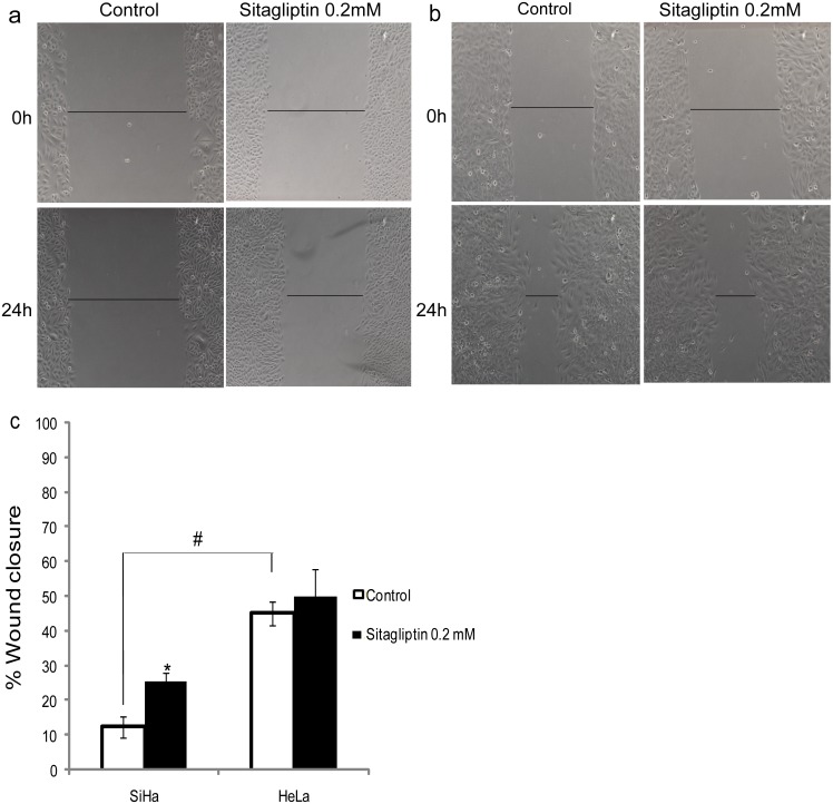
Fig 4. Cell migration using the Wound-healing assay.
SiHa (a) and HeLa (b) cervical cancer cells were analyzed 24 h after scratching in the absence (control) and presence of 0.2 mM sitagliptin phosphate. (c) Quantification of the wound closure of cervical cancer cells in the absence (control) and presence of sitagliptin phosphate. Results are mean values ± SD (n = 3). *Indicates statistical significance when the sitagliptin phosphate group was compared to the respective control group. #Indicates statistical significance when controls were compared (ANOVA followed by Tukey’s test, p ≤ 0.05).

