
Fig. 1.
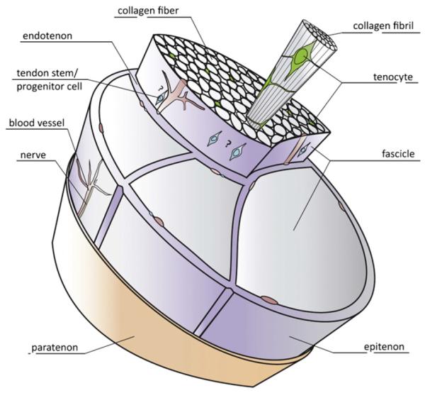
A schematic drawing of basic tendon structure. The collagen molecules are organized hierarchically in fibrils, fibers and fascicles. The cellular content is dominated by the tenocytes, which are terminally differentiated cells. Tendons contain stem and progenitor cell populations, whose exact location is still debated (therefore indicated with a?). Different sheets, endotenon and epitenon (loose connective tissues), and paratenon (fatty areolar tissue) are shown as well as blood vessels and nerves.
Based on [227].