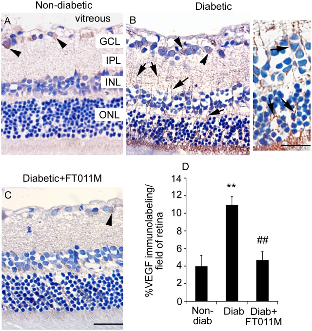
Fig 4. FT011M reduced VEGF immunolabeling in retina of Ren-2 rats diabetic for 8 weeks.
Non-diab, non-diabetic. Diab, diabetic. V, vehicle. Three-μm paraffin sections. GCL, ganglion cell layer. IPL, inner plexiform layer. INL, inner nuclear layer. ONL, outer nuclear layer. Counterstain, haematoxylin. (A). In non-diab + V, VEGF immunolabeling is occasionally detected in ganglion cells (arrowheads) in the GCL. (B) In diab + V, VEGF immunolabeling is increased compared to non-diab + V and detected in ganglion cells and Müller cell processes (arrows) extending throughout the retina. Inset showing higher powered image of Müller cell processes. (C) In diabetic rats treated with FT011M, VEGF immunolabeling was reduced to the level of non-diab + V. (A to C) Original magnification, 400X. Bar, 40 μm. Inset: Bar, 75 μm. (D) **P < 0.01 to non-diab + V. ##P < 0.01 to diab + V. N = 4 to 6 rats per group. Values are Mean ± SEM.

