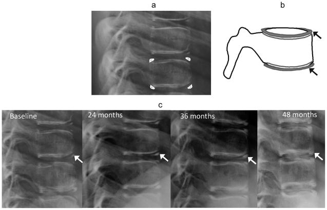
Fig. 1.
Ring apophyses. (a) Lateral radiograph ossification of ring apophyses in a 10 year-old girl on glucocorticoid treatment for chronic renal disease. (b) Corresponding diagram of development of ring apophyses at endplate margins (arrows). (c) Lateral radioography shows ossification and fusion of ring apophyses (arrows) over a four year period in the same girl

