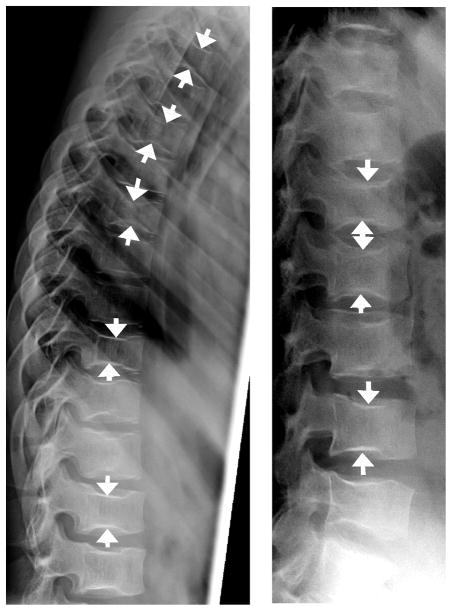
Fig. 12.
Sickle cell anemia. Lateral radiographs (a, b) show central endplate deformity at multiple levels in 13 year-old girl with sickle cell anemia, mimicking biconcave endplate compression fractures. Nearly all levels are involved to variable extent, with arrows showing some of the most prominent endplate depressions in this girl

