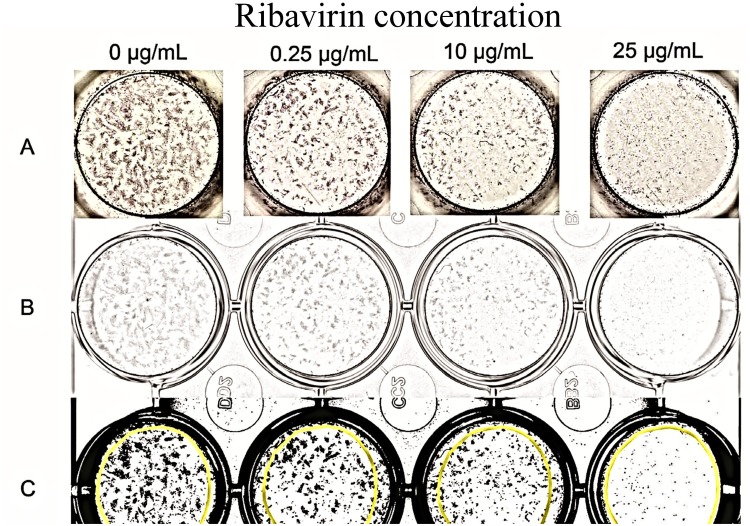
Fig 1. Digital image analysis to determine stained PaBV-4 positive cell foci and area of infected cells.
(A) Photograph after indirect immunocytochemistry of wells containing infected duck embryonic fibroblasts infected with PaBV-4 and incubated with or without ribavirin. PaBV-4 nucleoprotein detected as dense shades of grey. (B) Digital 8-bit grey-scale images of the same wells after digital scanning. (C) The same wells after 8-bit grey scale images were enhanced and converted to a binary image for analyses using ImageJ Analyze. The areas of N protein immunocomplex are in black. The area of interest that was analyzed is the same for each well and is within the superimposed yellow circles.

