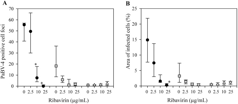
Fig 3. Anti-viral activity of Ribavirin on PaBV-4 in duck embryonic fibroblasts (DEF).
(A) PaBV-4 positive cell foci, assessed by indirect immunocytochemistry assay. (B) Percent area of infected cells, assessed by indirect immunocytochemistry assay. DEF infected with 64 ffu (black circle), 7 ffu of PaBV-4 (grey square), or no virus (white triangle), incubated with ribavirin for 5 days, and then incubated a further 5 days in the absence of ribavirin. Data presents as median ± 25th and 75th percentile. *, compared with no added ribavirin (P <0.05, ANOVA on ranks with Tukey test).

