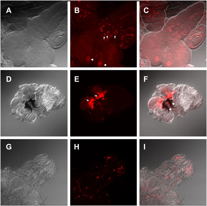
Fig 7. Expression pattern of the esg L4 enhancer trap.
(A-C) L4>UAS-GFP third instar larval central nervous system (CNS) and adjacent imaginal discs A) Differential interference contrast (DIC) microscopy image. (B) Confocal maximal projection of an immunohistochemistry against GFP. Arrows show the medial section of the thoracoabdominal ganglion esg expressing neurons and arrowheads show mushroom bodies. (C) Overlay of DIC and confocal images. (D-F) L4>UAS-GFP third instar larval inverted maxillary region that includes labial imaginal discs. (D) DIC microscopy image. (E) Confocal maximal projection of GFP fluorescence. Arrows show the expression domain in the proximal labial discs. Arrowheads show mandibles. (F) Overlay of (D) and (E). (G-I) L4>UAS-Stinger late pupal proboscis. (G) DIC microscopy image. (H) Confocal maximal projection of stringer fluorescence. (I) Overlay of (H) and (I). White dash-lines show the border of the CNS and labial imaginal discs, respectively.

