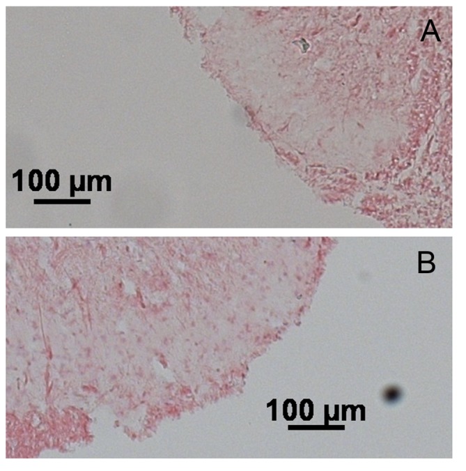
Fig 6. Representative images of mir-17-5p In situ hybridization in the dorsal horn of the spinal cord using the NBT-BCP detection system.

A) Spinal cord isolated from control rats. B) Spinal cord isolated from stressed rats. Increased mir-17-5p staining is observed in the dorsal horn. Scale bar is 100mM.
