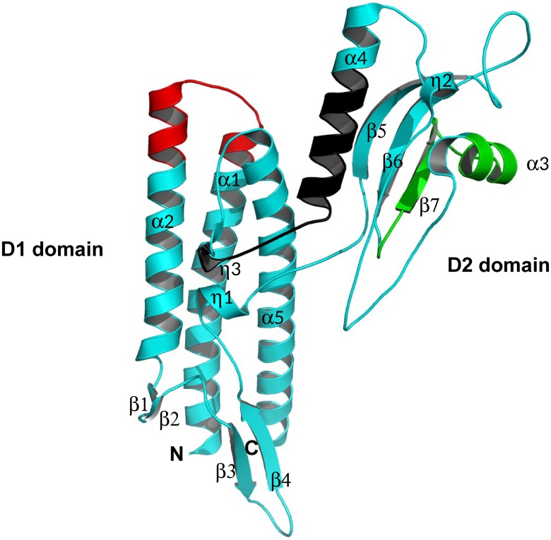
Fig 6. Domain organization and 3D crystal structure of FliCBp.
Schematic secondary structure ribbon representation of the crystal structure of FliCBp. The N- and C-termini are labeled as are the diverse secondary structural features with α-helices in cyan and β-strands in magenta. Peptides 96–111, 213–233 and 270–288 are highlighted in red, green and black, respectively. This figure was produced using MacPymol.

