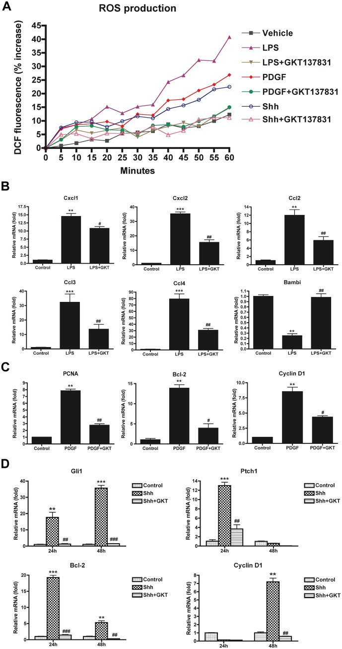
Fig 5. ROS production and gene expression were attenuated by GKT137831 in HSCs in response to LPS, PDGF and Shh stimulation.
(A) HSCs isolated from WT mice were loaded with H2DCFDA (10 μM) for 20 min. Cells were then washed and subsequently induced with LPS (100 ng/ml), PDGF (10 ng/ml) and Shh (1 μg/ml) in the presence or absence of GKT137831 (5 μM). ROS production was assessed by fluorescent signals quantified continuously for 60 min using a fluorometer. (B) Chemokine genes were attenuated by GKT137831 in HSCs in response to LPS. HSCs isolated from WT mice were pretreated with GKT137831 (20 μM) for 30 min, and then treated with LPS (100 ng/ml) for 6 h. Chemokine genes were analyzed by quantitative real-time PCR. **P < 0.01, ***P < 0.001 vs control; #P < 0.05, ##P < 0.01 vs LPS. (C) Proliferative genes were attenuated by GKT137831 in HSCs in response to PDGF. HSCs isolated from WT mice were treated with PDGF (10 ng/ml) in the presence or absence of GKT137831 (20 μM) for 24 h. Proliferative genes were analyzed by quantitative real-time PCR. **P < 0.01 vs control; #P < 0.05, ##P < 0.01 vs PDGF. (D) Hedgehog genes were attenuated by GKT137831 in HSCs in response to hedgehog ligand Shh. HSCs isolated from WT mice were treated with Shh (1 μg/ml) in the presence or absence of GKT137831 (20 μM) for 24 and 48 h. Hedgehog genes were analyzed by quantitative real-time PCR. HPRT was used as an internal control. **P < 0.01, ***P < 0.001 vs control; ##P < 0.01, ###P < 0.001 vs Shh.

