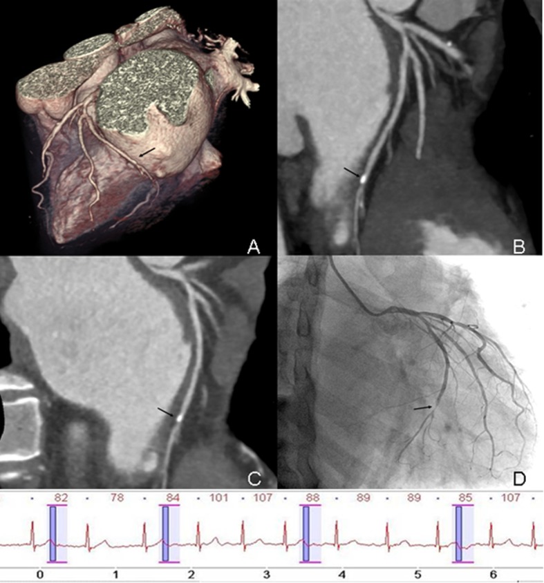
Fig 3. Prospectively ECG-triggered dual-source CCTA of a 60-year-old man with AF.

Mean HR was 86 bpm (range, 60–107 bpm). Images reconstructed at 240 ms after R wave. Volume-rendered (A), maximum intensity projection (B) and curved multiplanar reformation (C) image of LCX (black arrow) show the vessel lumen was overlapped by calcified plaque in the distal segment. Conventional coronary angiogram (D) shows significant stenosis (>50%) in distal segment of LCX (black arrow). The ECG information was recorded during data acquisition.
