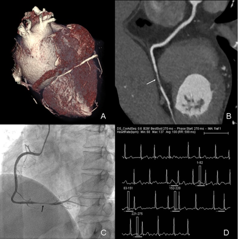
Fig 4. Prospectively ECG-triggered dual-source CCTA of a 57-year-old woman with AF.

Mean HR was 100 bpm (range, 68–137 bpm). Images reconstructed at 270 msec after R wave. Volume-rendered (A) and curved multiplanar reformation (B) images of RCA (white arrow) show atherosclerosis lesion (stenosis>50%) in distal segment. Conventional coronary angiogram (C) shows significant stenosis (>50%) in distal segment of RCA (black arrow). The ECG information (D) was recorded during data acquisition.
