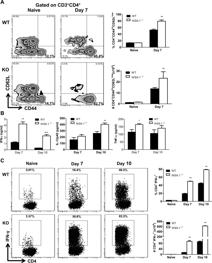
Fig 5. Enhanced activation of CD4+ T cells and elevated production of inflammatory cytokines in the liver of IL-27R-/- (WSX-1-/-) mice infected with T. congolense.
(A) The frequency (left and upper right) and the absolute number (lower right) of activated CD4+ T cells (CD44hiCD62Llow) derived from the liver of IL-27R-/- and wild-type mice (n = 3) on day 0 and 7 after infection with T. congolense. (B) Production of IFN-γ, IL-12p40, and TNF-α in the supernatant fluids of cultured liver leukocytes purified from IL-27R-/- and wild-type mice (n = 4) on day 7 and 10 after infection with T. congolense. (C) The frequency (left and upper right) and the absolute number (lower right) of IFN-γ-producing CD4+ T cells derived from the liver of IL-27R-/- and wild-type mice (n = 3) on day 0, 7 and 10 after infection with T. congolense following 12 h in vitro restimulation with Cell Stimulation Cocktail (containing PMA, ionomycin, and protein transport inhibitors). Data are presented as the mean ± SEM. The results presented are representative of 2–3 separate experiments.

