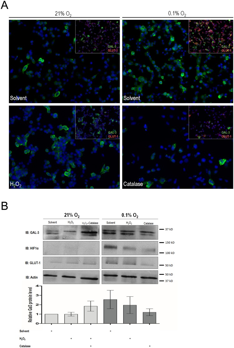
Fig 2. Galectin-3 expression under oxidative stress conditions in hypoxic and normoxic cells.
(A) Galectin-3 subcellular expression was assessed by double-labelling immunofluorescence. Representative galectin-3 cell expression shows that despite galectin-3 increase under hypoxia, in the presence of catalase this was not verified. GLUT-1, included as a well-known target of hypoxic conditions, was also not increased in the presence of catalase. (B) To quantitatively evaluate galectin-3 expression under different oxidative stress conditions western blot analyses were performed. Relative intensity of the indicated protein level bands normalizes to actin were measured. In hypoxic conditions, in the presence of catalase, increased expression of galectin-3 was not observed in CMT-U27 cells. Galectin-3 expression, was kept, both in levels and quantity, similar to that seen in normoxic cells. However, in normoxia, catalase itself appears to increase the expression of galectin-3. In normoxic conditions, hydrogen peroxide treatment induced an apparent increase of a heavier molecular weight form of galectin-3 but had no additive effect on the increase of galectin-3 under hypoxia.

