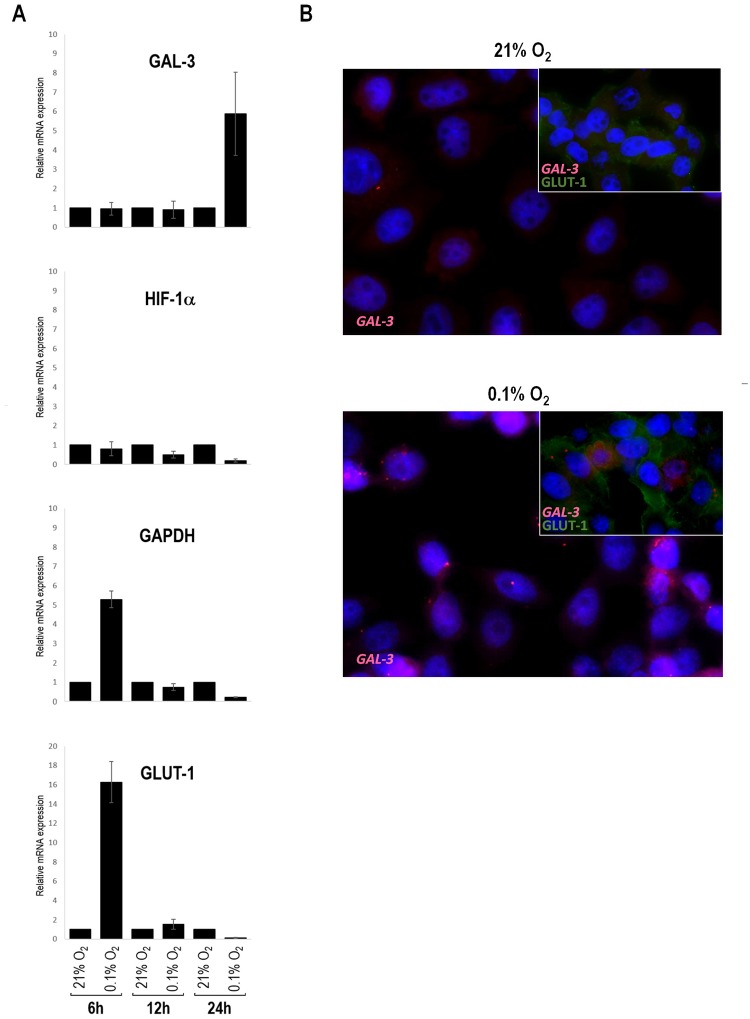
Fig 4. Galectin-3 mRNA expression under hypoxic conditions.
(A) mRNA extracted from malignant CMT-U27 cell line was converted to cDNA and analyzed by real-time polymerase chain reaction (PCR) to assess quantitatively galectin-3 expression in CMT-U27 cells under hypoxic conditions. An increase in galectin-3 mRNA was observed only upon 24 hours of hypoxia treatment. No differences were seen in the transcription of HIF-1α, however the transcription of GAPDH and GLUT-1 seemed to early respond to hypoxia. cDNA contents were normalized on the basis of predetermined levels of 18S. (B) Representative mRNA expression of galectin-3, was visualized using a set of Stellaris RNA fluorescence in situ hybridization probes. GLUT-1 (green color) was used as a hypoxia control. Blue color shows the nucleus stained by DAPI. CMT-U27 cells exposed for 24 hours to hypoxia presented intense red FISH signals that are predominantly located in the cytoplasm and reflect the galectin-3 mRNA. Each spot corresponds to a single mRNA molecule.

