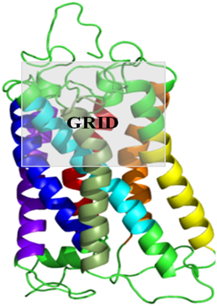
Fig 1. GRID selected for Induced Fit Docking.
Induced Fit docking protocol was standardized using the experimental data available on mouse ORs that responds to eugenol (mOR-EG). Different grid parameters and constraints were used to standardize the protocol as shown in Table 2. The use of the upper half of the receptor facing the extracellular milieu gave the best score for eugenol binding as compared to the other parameters. Thus similar grid parameters were used for all the IFD runs. The receptor TM helices 1–7 are coloured in VIBGYOR colour (Violet, Indigo, Blue, Green, Yellow, Orange and Red). Figure has been generated using PYMOL (The PyMOL Molecular Graphics System, Version 1.5.0.4 Schrödinger, LLC).

