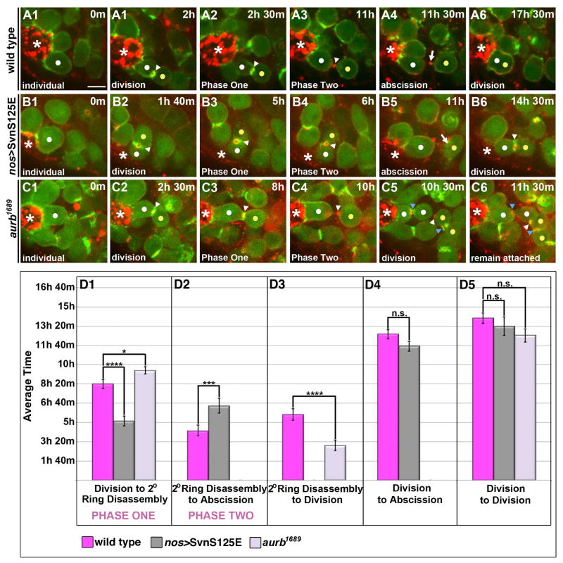
Figure 5. AurB/Svn activity temporally regulates the transition between Phases One and Two of delay.
(A1–C6) Time lapse imaging of GSC division cycle; ABD-moeGFP and Myo-mCherry. Each panel is a 3–5 Z plane projection. m=min. h=hour. White dot=GSC. Yellow dot=Gb. White arrowhead=“original” IC bridge. Blue arrowhead=“new” IC bridge. Arrow=midbody remnant. *=Hub. Scale bar=5 microns
(A1–A6) Progression through two rounds of mitosis in a wild type GSC (same GSC images shown in Fig. 1).
(B1–B6) Progression through two rounds of mitosis in a GSC expressing SvnS125E that completes abscission.
(C1–C6) Progression through two rounds of mitosis in a GSC depleted of AurB that fails to complete abscission. The group of four cells (C5–C6) remained connected and associated with the hub for the remainder of our imaging (1–3 hrs after second division). An F-actin ring was eventually reestablished at the “original” IC bridge (white arrowhead).
(D1–D5) Quantification of timing of indicated phases. The same GSC-Gb pairs were followed through each stage reported. (D1) Expression of activated Svn led to precocious exit from Phase One (p=3.6 × 10⁻⁴) while depletion of Aurb had the reciprocal effect of prolonging Phase One (p=0.03). (D2) SvnS125E-expressing pairs spent a significantly longer time, on average, in Phase Two compared with controls (p=9.0 × 10⁻³). This likely reflects some conservation of the described role for AurB/Svn activity in delaying abscission. (D3) As GSCs depleted of AurB did not abscise, we instead quantified the time from secondary ring disassembly to division. Loss of AurB activity led to a statistically significant decrease in the average time from secondary ring disassembly to division compared with controls (p=6.7 × 10⁻⁴). (D4) The average time from division to abscission was not significantly different in SvnS125E-expressing pairs compared with controls (p=0.11) despite their precocious exit from Phase One. This is due to the increased length of Phase Two, which, as described above, reflects conservation of a role for the CPC in directly regulating abscission timing. (D5) GSCs followed through two rounds of mitosis revealed no significant difference in the cell cycle rate of GSCs expressing SvnS125E (p=0.43) or depleted of AurB (p=0.05) compared with controls. Data are represented as mean +/− SEM.

