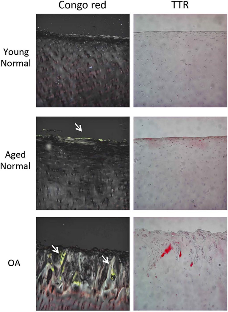
Figure 1. Amyloid and TTR deposition in human young normal, aged normal and OA cartilage.
Representative images of Congo red staining and Immunohistochemistry for human TTR in young normal, aged normal, and OA cartilage. Arrows indicate green staining of amyloid under polarized light. Images are 10× magnification.

