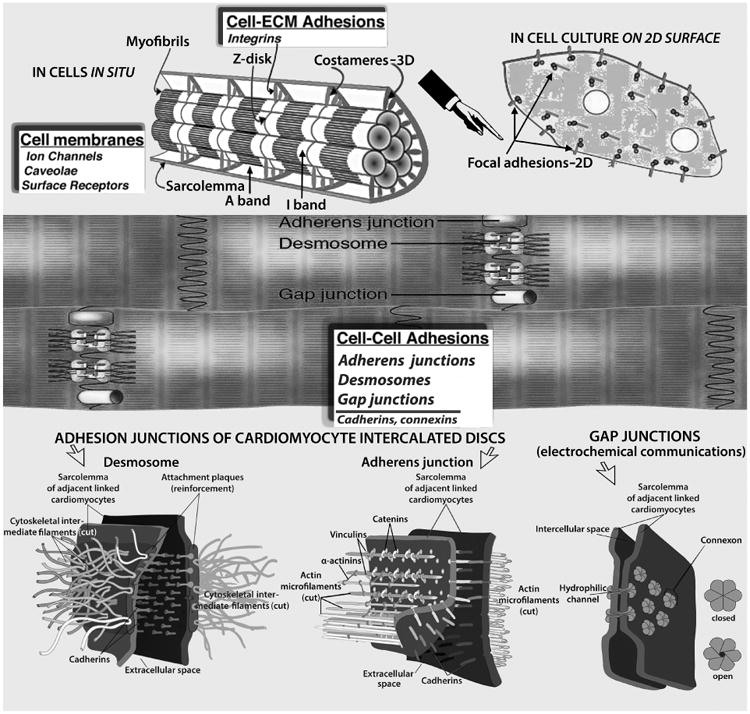
Figure 2.

Myocardial cell junctions can be divided into two types: those that link cells to the ECM (costameres and focal adhesions, Top), and intercellular junctions (gap, adherens, and desmosomes, Middle & Bottom) that link cells together directly. Radially arranged integrins, and other specialized proteins, constitute physical links between the Z-disk and sarcolemma; they transmit contractile forces from sarcomeres across the sarcolemma laterally to the extracellular matrix and ultimately to neighboring cardiomyocytes. The desmosome, like the adherens junction, comprises calcium ion-dependent cell adhesion molecules that interact with similar molecules in the adjacent cell. Adherens junctions and focal adhesions not only tether cells together or to the ECM, but they also transduce signals into and out of the cell, influencing a variety of cellular epigenetic responses, notably to flow-associated forces and deformations. The basic building block of the gap junction is the connexin subunit. Six of these in each of the membranes of two adjacent cells come together to form a connexon that interacts with a comparable hexamer in the other cell resulting in formation of a channel, which allows cytoplasmic communication between the cells. Myocardial cell membranes encompass ion channels, surface receptors and caveolae—not shown. (See discussions in text). Costamere diagram adapted, modified, from Ervasti JM [31].
