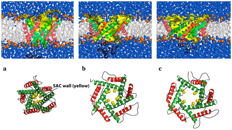
Figure 3.

Schematic of numerical simulation of the gating process of a sarcolemmal mechanosensitive ion channel. Putting the membrane under tension, the stretch-activated channels (SAC) undergo significant conformational changes in accordance with an iris-like dilation mechanism, reaching a conducting state on a microsecond timescale. Diagrammed is a channel with (a) small, (b) intermediate, and (c) large opening under the action of rising membrane tension. Top: side view showing the lipid bilayer (blue denotes an aqueous environment, yellow represents the SAC wall). Bottom: End view showing the simulated membrane pore. Reproduced, slightly modified, with permission from Yefimov S, et al [77].
