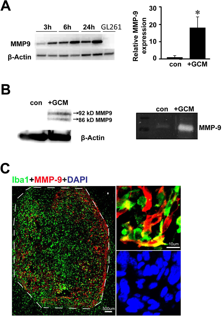
Fig. 1. Microglial cells are upregulating MMP-9 when associated with gliomas.
(A) MMP-9 gene expression in microglia stimulated with GCM for 3h, 6h and 24h was analyzed by RT-PCR (left). GL261 cells were also analyzed for MMP-9 expression. β-actin serves as a loading control. Microglial MMP-9 expression was quantified with qRT-PCR after 24h stimulation with GCM (right). Bars represent the mean±s.e.m. from 3 independent experiments. (B) Western blot from both cell lysate (left panel) and gelatin zymography from supernatant (right panel) showed MMP-9 induction in microglia upon GCM stimulation for 24h. (C) Mouse brains injected with GL261 glioma cells were stained for microglial marker Iba-1(green) and for MMP-9 (red). In the tumor free area the level of MMP-9 was low (left, out of the dash line) while within the tumor MMP-9 is expressed mainly in Iba-1 positive cells (right).

