
Figure 2. Immunohistochemistry of Gal-1 expression in mouse testis sections.
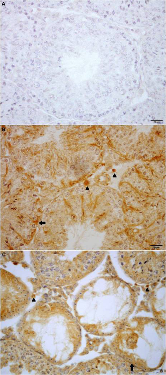
Photomicrographs of paraffin-embedded sections of normal (B) and experimental (C) adult testes immunostained with Gal-1 antibody. Strong Gal-1 immunoreactivity can be observed within the seminiferous tubules associated with both Sertoli cells (arrow) and differentiating germ cells and also in Leydig cells (arrowhead) located in the interstitial spaces. Omission of primary anti-Gal-1 antibody was used as a negative control (A).