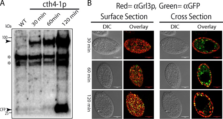
FIG 2.
Expression and localization of CFP-tagged Cth4p. (A) Western blot, probed with anti-GFP MAb that cross-reacts with CFP, of cells expressing CFP-tagged Cth4p (cth4-1p). cth4-1p expression was induced for 30, 60, and 120 min with 1 μg/ml of CdCl2. At these times, cell lysates were prepared by trichloroacetic acid (TCA) precipitation and then solubilized in SDS-PAGE sample buffer. Proteins fractions were separated by 4 to 20% SDS-PAGE and transferred to polyvinylidene difluoride (PVDF). Molecular mass standards are shown on the left. The specific bands recognized by the antibody, indicated by arrowheads, are of the size expected for full-length Cth4p-CFP (94 kDa) and monomeric CFP. At 30 and 60 min of induction, only full-length Cth4p-CFP was detected, whereas monomeric CFP was also detected after 120 min. Asterisks indicate nonspecific cross-reactive species. (B) Using the same cultures as for panel A, cells at 30, 60, and 120 min were fixed and immunolabeled with mouse MAb 5E9 to localize mucocyst protein Grl3p and rabbit anti-GFP to localize the Cth4p-CFP fusion. The fluorescent puncta at the cell surface, which appear as elongated vesicles in cross section, correspond to the array of docked mucocysts. Scale bars = 10 μm.

