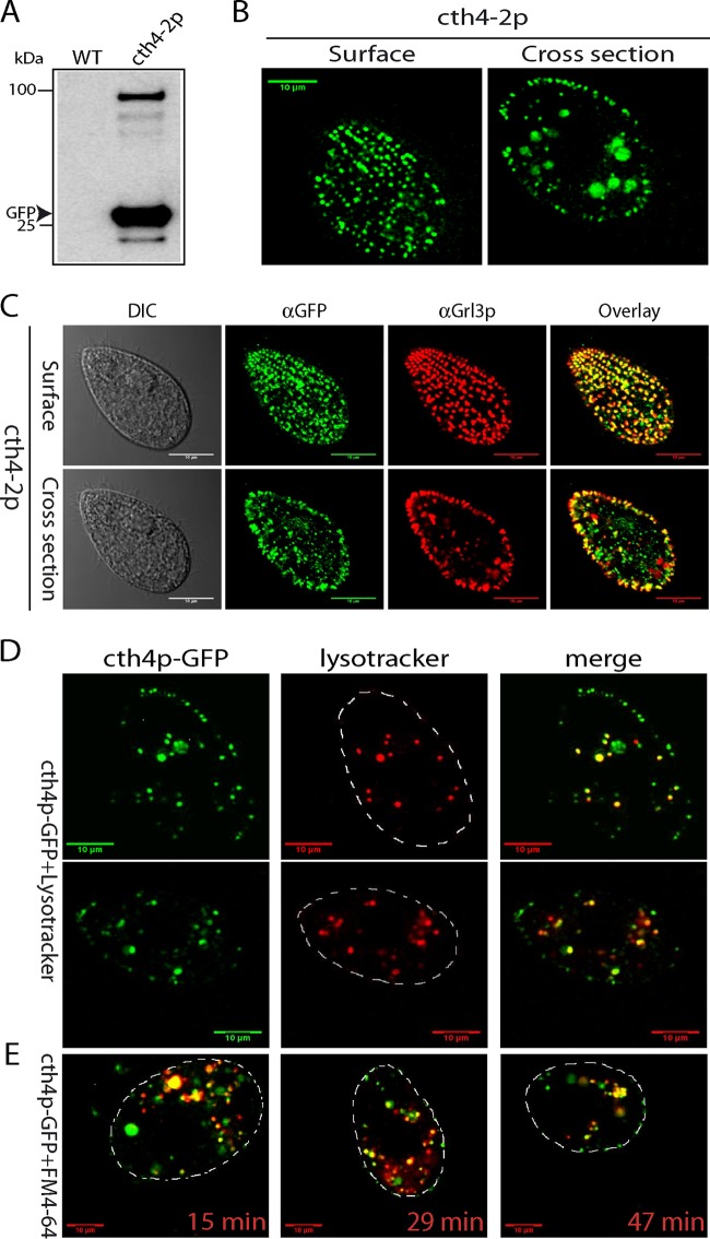
FIG 3.
Localization of endogenous Cth4p-GFP. (A) Expression of cth4-2: GFP-tagged Cth4p from the endogenous CTH4 locus, under the control of its native promoter. cth4-2p was immunoprecipitated from detergent lysates using polyclonal rabbit anti-GFP antiserum, and immunoprecipitates were Western blotted with monoclonal anti-GFP Ab. One immunoreactive band is of the size expected for the Cth4p-GFP fusion, while a second is the size expected for monomeric GFP. (B) Live immobilized cells expressing cth4-2p were imaged to capture cell surface and cross sections and show the expected array of docked mucocysts as well as intracellular puncta. (C) Immunostaining of fixed cells to simultaneously localize mucocyst protein Grl3p and cth4-2p. There is extensive colocalization in docked mucocysts, while some puncta deeper in the cytoplasm are positive for cth4-2p but not Grl3p. (D) Cells were incubated for 5 min with 200 nM Lysotracker, and live images were captured within 30 min. Optical sections shown are cell cross sections. (E) Cells were incubated for 5 min with 5 μM FM4-64, an endocytic tracer and then pelleted and resuspended in tracer-free medium. The times shown represent minutes after resuspension. Scale bars = 10 μm.

