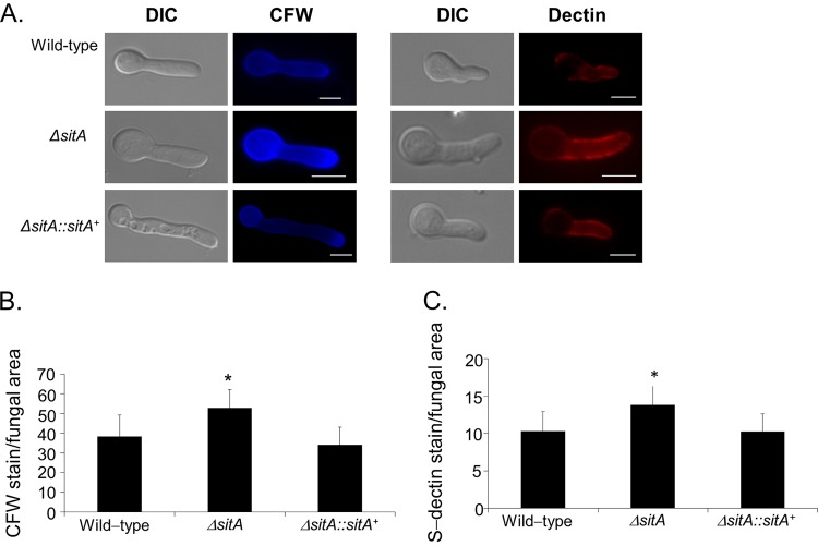
FIG 4.
Detection of the β-(1,3)-glucan and chitin contents on the cell surface. Conidia were cultured in liquid medium to the hyphal stage, UV killed, and stained with soluble dectin-1 or calcofluor white (A) to detect the content of exposed chitin (B) or β-glucan (C), respectively. The intensity of staining was calculated by averaging the amount of staining to the total area of each fungal cell using ImageJ software. The experiments were performed in triplicate, and the results are displayed as arbitrary units (mean values with standard errors; * = P < 0.05 by t tests). Bars, 5 μm. DIC, differential interference contrast.

