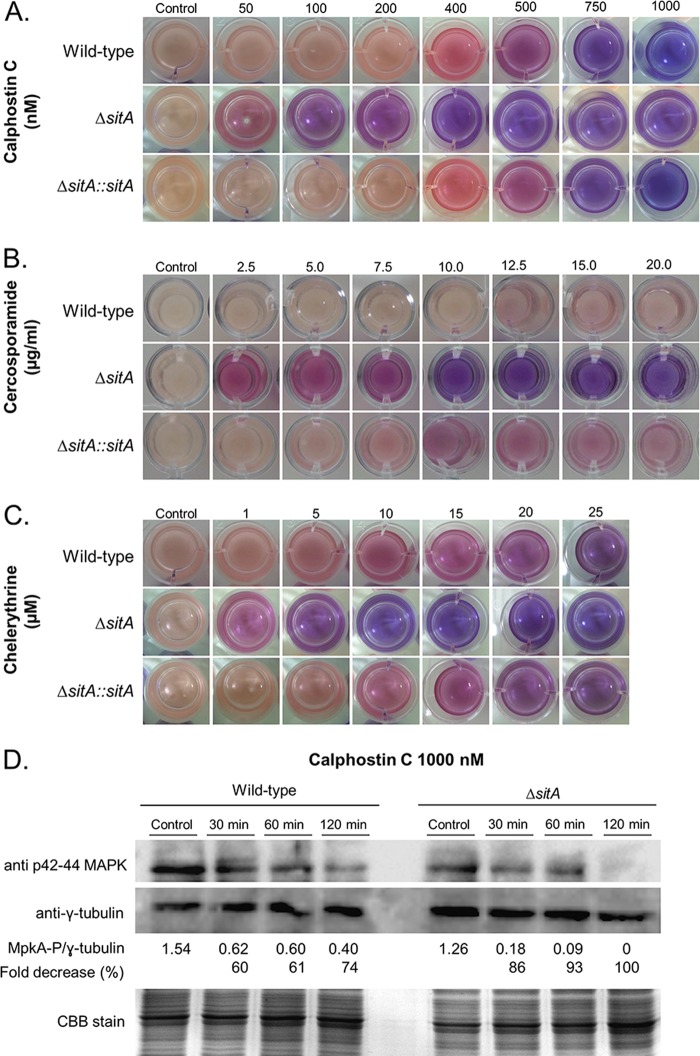
FIG 8.
The ΔsitA strain has reduced protein kinase activity. (A to C) Viability of the germlings of the wild-type, ΔsitA, and ΔsitA::sitA+ strains grown in the absence or presence of calphostin C, cercosporamide, and chelerythrine as shown by the viability indicator alamarBlue. The germlings are less viable when the indicator shows intensely blue colonies, indicating decreased mitochondrial activity. Pink and red indicate viability. (D) Western blot for MpkA phosphorylation. The wild-type and ΔsitA strains were grown for 16 h at 37°C, and then mycelia were transferred to fresh medium with calphostin C for 30, 60, and 120 min. Anti-phospho-p44/42 MAPK antibody directed against phosphorylated MpkA was used to detect the phosphorylation of MpkA. Anti-γ-tubulin antibody was used as a control for loading. A CBB-stained gel is shown as an additional loading control. The signal intensities were quantified using Image J software by dividing the intensity of MpkA-P by that of γ-tubulin.

