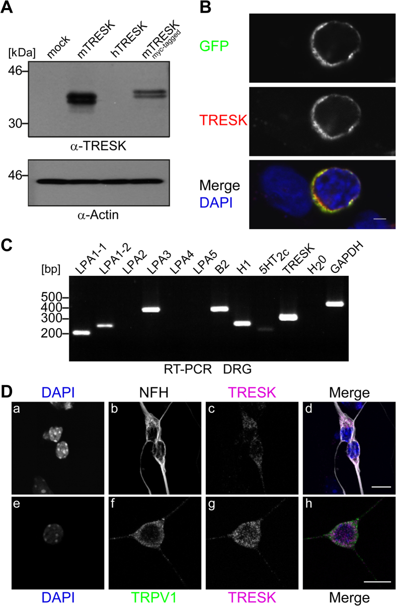
Figure 1. Expression profile of LPA receptors and immunodetection of TRESK channel in DRG neurons.
(A) Whole cell extracts of HEK-293 cells transfected with pcDNA3 (mock), mTRESK, hTRESK and myc-mTRESK plasmid DNA (as indicated) were immunoblotted and analysed with polyclonal TRESK antibody (upper panel) or anti-Actin antibody as loading control (lower panel). Extracts of cells transfected with mTRESK or myc-mTRESK displayed double bands with the expected molecular weight indicating high specificity for mouse TRESK of the novel antibody. (B) HEK-293 cells were transfected with mTRESK-eGFP fusion construct and analysed by confocal microscopy. Only transfected cells (GFP in upper panel) were found to have membrane reactivity with TRESK antibodies (middle panel). Lower panel, merge displayed co-reactivity of the antibody with GFP signal. DAPI staining in blue revealed also non-transfected cells. Scale bar 2 μm. (C) Expression of mTRESK, and a selection of Gq-coupled receptors (as indicated) was monitored in adult DRGs by RT-PCR. Primers for GAPDH were used as a positive control and H2O was applied as a negative control in reverse transcription. (D) Immunocytochemistry of cultured DRG neurons from E13.5 mouse embryos probed with antibodies against TRESK (c, g), NFH (b), TRPV1 (f) and DAPI (a, e). Images a-c and e-g are merged in panel d and h, respectively. Merge in d documented that only NFH-stained neurons (grey) displayed TRESK reactivity (magenta); DAPI-stained, non-neuronal cells (a, upper and lower edge) displayed hardly any TRESK signal (c). Neuronal co-expression of TRESK (magenta) and TRPV1 (green) was documented in merge of panel h (out of 29 TRESK-positive cells 18 were also positive for TRPV1). Fused z-stacks of confocal images are shown. Scale bar 10 μm.

