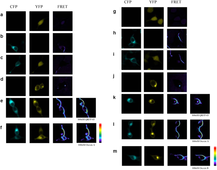
Figure 5. FRET imaging of constitutive GPR103 and OX1R/OX2R heteromeric interactions in living cells.
GPR103-CFP and OX1R-YFP were expressed individually or co-expressed in HEK293T cells: YFP (a) GPR103-CFP (b) GPR103-CFP and YFP (c) OX1R-YFP (d) GPR103-CFP and OX1R-YFP (e,f) Upon stimulation with 100 nM QRFP-43 (e) or OXA (f) there is significant change in FRET. GPR103-CFP and OX2R-YFP were expressed individually or co-expressed in HEK293T cells: YFP (g) GPR103-CFP (h) GPR103-CFP and YFP (i) OX2R-YFP (j) GPR103-CFP and OX2R-YFP (k–m) FRET was detectable upon stimulation with QRFP (k) OXA (l), OXB (m; all at 100 nM). Lowest FRET intensity is in purple/black and the highest in red.

