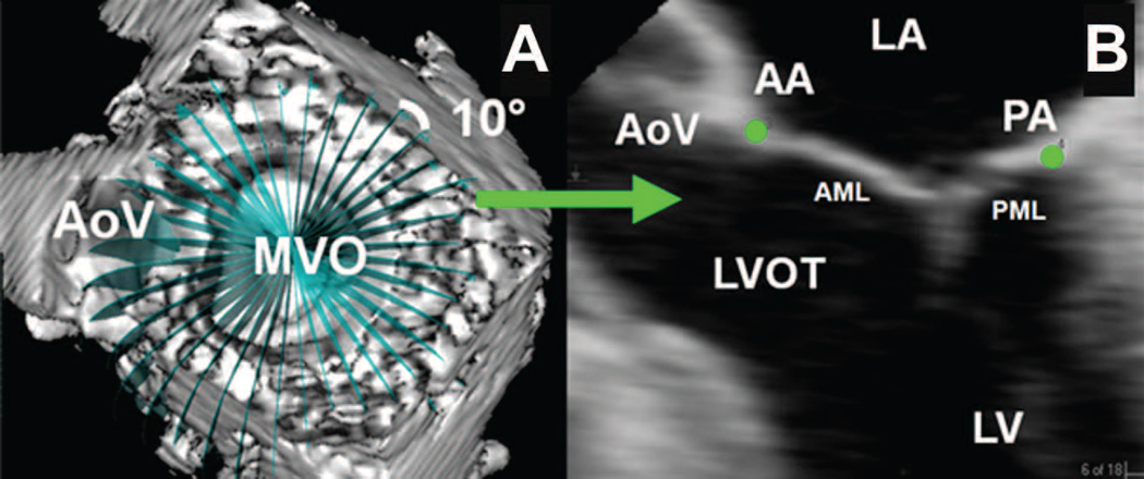
Figure 1. Annular Segmentation Technique.
Panel A demonstrates a view of the mitral valve where the selected short axis plane coincides with the plane of the mitral valve. The aorta valve (AoV) and mitral valve orifice (MVO) are indicated. A rotational template consisting of 18 long axis planes evenly spaced at 10° increments and centered at the geometric center of the mitral valve are constructed. Panel B demonstrates a single long axis view (0 degrees on the rotational template of Panel A) of the heart. The left ventricle (LV), left ventricular outflow tract (LVOT), anterior (AML) and posterior (PML) mitral leaflets, left atrium (LA), aortic valve (AoV), anterior (AA) and posterior (PA) annular points have been marked. Note that in this orientation, the negative z-axis (for purposes or annular height and coaptation height calculations) extends towards the apex while the positive z-axis extends towards the left atrium.

