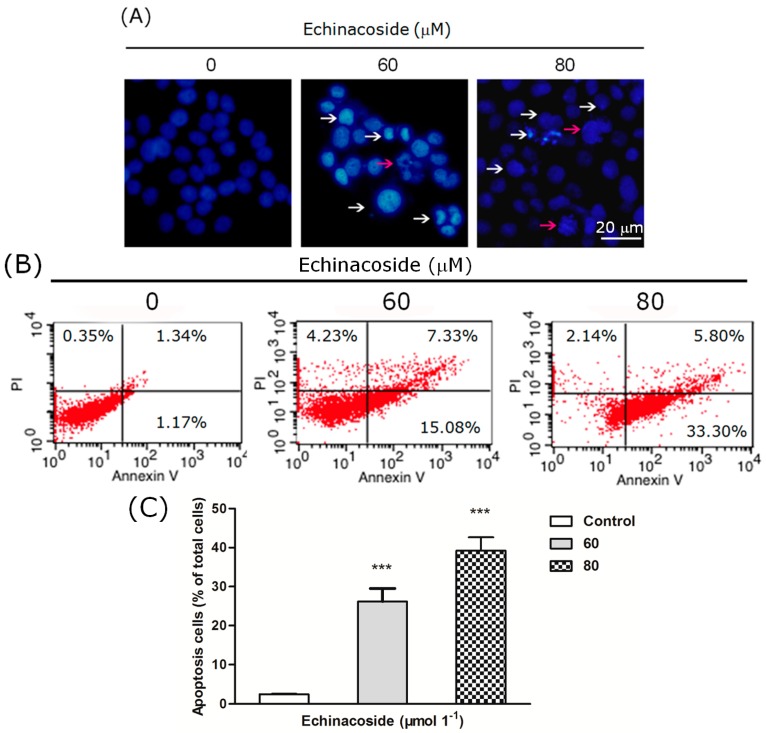
Figure 3.
(A) Images of 4,6-diamidino-2-phenylindole (DAPI)-stained cells: Cells were treated by 0, 60 and 80 μM Echinacoside for 24 h, some apoptotic cells were marked by white and red arrows (scale bar = 20 μm); (B) Flow cytometry analysis of apoptosis: Cells were stained by Annexin V-FITC and Propidium Iodide, and analyzed by the FACSCalibur flow cytometer and the Cell Quest software; (C) Quantification of apoptotic cells from three independent experiments, data were analyzed using the GraphPad Prism software (*** p < 0.001 vs. vehicle control).

