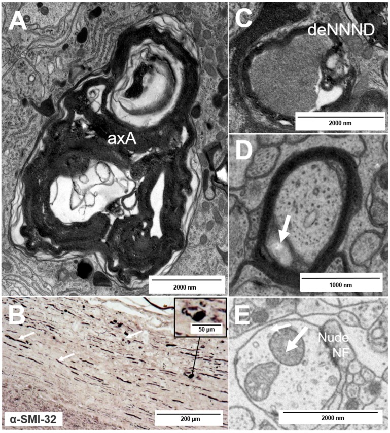
Figure 4.
Types of nerve fiber pathology: (A) axolytic axons (axA); (B) axonal transection as observed after staining for hypophosphorylated neurofilaments (arrows) with typical “ovoid” formation (inset); and (C–E) fine and early nerve fiber pathology: a decrease in the nearest neighbor neurofilament distance (C, deNNND), enlargement of the inner tongue (D, arrow) and a nude nerve fiber (nudeNF) with enlarged mitochondria (E, arrow).

