Figure 4.
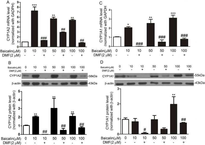
Baicalin increased CYP1A1 and CYP1A2 expression at the mRNA and protein level in Fa2N-4 cells. Cells were treated with baicalin (0–100 μM) for 24 h in the presence or absence of 3ʹ,4ʹ-dimethoxyflavone (DMF) (2 μM), a specific antagonist of AhR. mRNA level of CYP1A2 was determined with real-time PCR and data were normalized to the expression of GAPDH (A); Protein expression of CYP1A2 was assessed through Western blot and data were normalized to that of β-actin (B); mRNA level of CYP1A1 was determined with real-time PCR and data were normalized to the expression of GAPDH (C); Protein expression of CYP1A1 was assessed through Western blot and data were normalized to that of β-actin (D). Data are expressed as the mean ± SD (n = 5). * p < 0.05, ** p < 0.01, *** p < 0.001 versus the control group; # p < 0.05, ## p < 0.01, ### p < 0.001 versus the baicalin only group.

