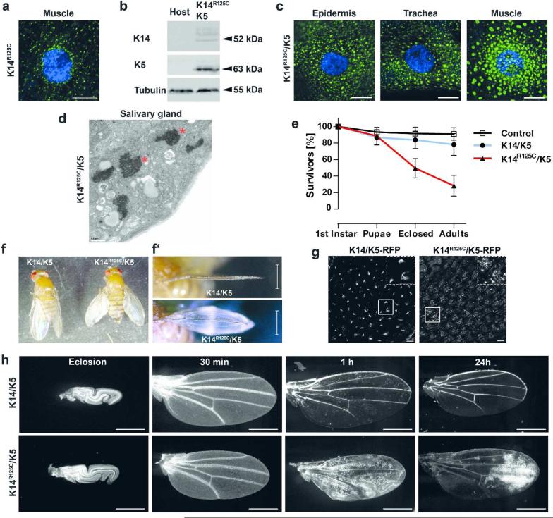
Figure 2. The human disease-causing mutant K14R125C causes wing blistering.
a) Confocal immunofluorescence image of muscle tissue from third instar larvae expressing K14R125C (green) using Mef2-GAL4 muscle-specific driver. DAPI-stained DNA is indicated by blue. b) Western analysis of total protein extracts of adult flies expressing K14R125C/K5 using Act5C-GAL4 and extracts from the host stock, which does not express keratins. Full size image of the western is shown in Figure S1. c) Confocal immunofluorescence images of third instar larval tissues expressing K14R125C (green) and K5. K14R125C/K5 were expressed in epidermis and trachea by Act5C-GAL4 or muscle specific by Mef2-GAL4. DAPI-stained DNA is indicated by blue. d) Electron microscopy images of salivary glands of third instar larvae expressing K14R125C/K5 by the salivary gland specific by Sgs3-GAL4 driver. Asterisks indicate keratin aggregates. e) Survival curves of flies expressing wt K14/K5 and K14R125C/K5 keratins by Act5C-GAL4. Adults were analyzed 3 to 4 days after eclosion from the pupal case. Mean ± SD, n≥6. f) Image of adult flies expressing wt or mutant K14 in combination with K5 using Act5C-GAL4. Frontal views of the wings are shown in f’). g) Expression of K14/K5-RFP or K14R125C/K5-RFP in wings immediately after unfolding, using Act5C-GAL4. RFP epifluorescence signal was recorded 5 to 10 min after unfolding. Dashed boxes are magnification of smaller boxes. h) Intervein cell clearance in K14/K5 or K14R125C/K5 expressing flies using Act5C-GAL4,UAS-GFP. GFP epifluorescence signal in wings were recorded at eclosion and at indicated time points after wing unfolding. Scale bars: a/c/g 10 μm; d 0.5 μm; f’ and h 500 μm

