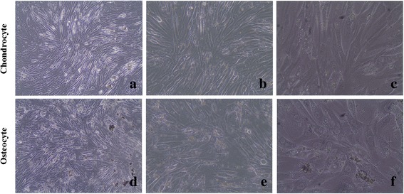
Fig. 2.

Inverted light microscopy images of chondrocytes and osteocytes cultures (a, b, and c are ×10, ×20, and ×40 magnification of approximately 97 % confluent chondrocytes, respectively; d, e, and f are ×10, ×20, and ×40 magnification of approximately 91 % confluent osteocytes, respectively)
