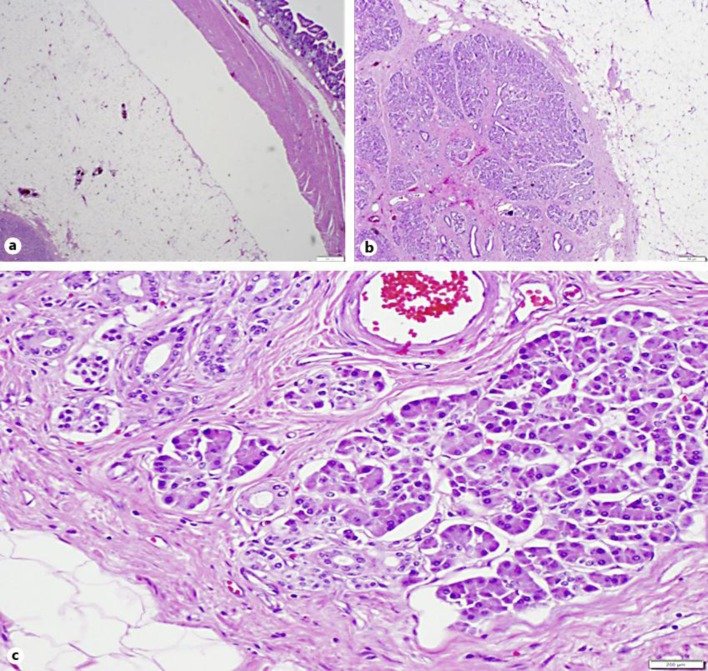
Fig. 3.
Histological sections (H&E) of the terminal ileum ‘tumor’. a Low-power (×2) cross-section. The submucosal lipoma, the centrally located benign pancreatic tissue and the overlying circular small intestinal mucosa are well illustrated. b, c Low-power (b, ×4) and medium-power (c, ×10) H&E section revealed the presence of pancreatic acini and ducts, surrounded by mature adipose tissue of the lipoma. No dysplasia or atypia is seen.

