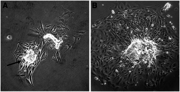
Fig. 1.

Representative images of pig mammary organoids collected after enzymatic dissociation of mammary gland tissue from a non-pregnant female. a A cluster of epithelial cells (closed arrowhead) and piece of duct (open arrowhead) with surrounding outgrowth 24 h after dissociation and plating. b An organoid with typical outgrowth 48 h after dissociation. Scale bar = 100 μm
