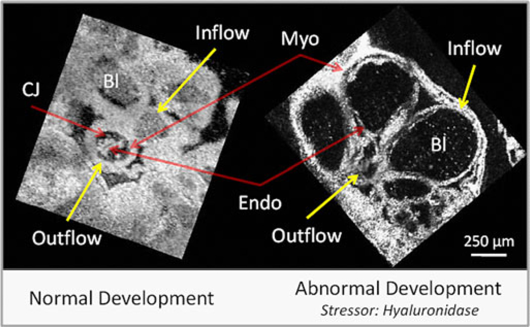
Fig. 6.
Normal versus abnormal heart development. En face slices from 4-D image sets (90 volumes/heartbeat). Hyaluronidase degrades the proteoglycan component of the cardiac jelly found between the myocardium and the endocardium. From the images, it is clear that there is a reduction of blood cells and the myocardial walls are thinner in the perturbed heart. Also, the maximum wall velocity between the inflow (2.4 mm/s) and outflow (0.6 mm/s) tract varies significantly in normal development, while the perturbed heart did not have a higher maximum velocity in the inflow. The decreased inflow wall velocity suggests that the cardiac jelly may play an important role in regulating cardiac conduction. CJ—cardiac jelly; Myo—myocardium; Endo—endocardium; and Bl—blood.

