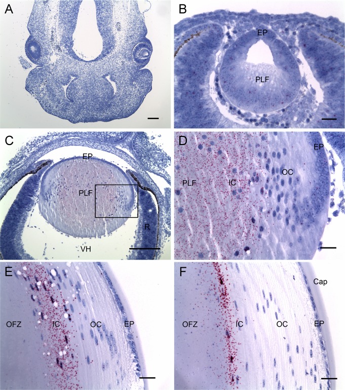
Figure 4.
In situ hybridization analysis of Livin mRNA expression in the developing mouse eye. (A) At day 12.5 of embryonic development, Livin expression (red dots) is undetectable in the embryonic head. (B) The lens, at this stage a hollow vesicle, is composed of epithelial cells (EP) and primary lens fibers (PLF) in the process of elongation. Livin expression in the E12.5 lens is barely above background. (C, D) At day 16.5 of embryonic development, Livin is expressed strongly by primary fiber cells located in the center of the lens. The boxed region in (C) is shown at higher magnification in (D). Livin expression is undetectable elsewhere in the eye. (E) At postnatal day 30, Livin expression is restricted to nucleated IC fiber cells bordering the central OFZ. (F) At age 6 months, Livin expression is restricted to a thin layer of fiber cells adjacent to the OFZ. Cap, capsule; R, retina; VH, vitreous humor. Scale bars: (A, C) 200 μm, (B, D–F) 25 μm.

